Scoliosis & Spinal Deformity
Comprehensive management of adult and adolescent scoliosis, kyphosis, flatback deformity, and post-surgical deformity. Conservative bracing to complex multi-level correction - tailored to your curve, your age, and your goals.
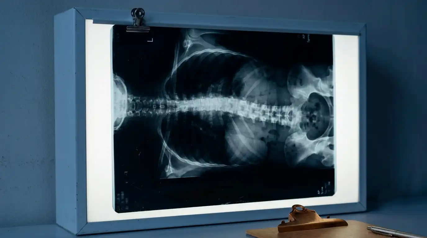
The most common form - curved spine without a known structural cause, typically identified during puberty.
Develops as disc and facet joints degenerate asymmetrically, causing progressive spinal curvature.
Abnormal forward or flattened curvature of the thoracic or lumbar spine, each requiring a distinct approach.
Deformity developing after previous spinal surgery - among the most technically demanding to correct.
Spinal curves caused by vertebral malformations present at birth, requiring early identification and monitoring.
Associated with cerebral palsy, muscular dystrophy, spina bifida, or spinal cord injury. Curves tend to be large and progressive.
Not every scoliosis patient needs surgery. The decision depends on curve magnitude (Cobb angle), rate of progression, patient age, skeletal maturity, symptoms, and quality of life impact.
We provide the full spectrum: structured observation programmes, custom bracing for adolescents, physiotherapy-based Schroth method for functional improvement, and surgical correction when indicated.
When surgery is needed, we use modern techniques including pedicle screw fixation, osteotomies, and minimally invasive approaches - with intraoperative neuromonitoring throughout.
Curves <20°, or slow-progressing curves in skeletally mature patients - monitored with periodic X-rays.
Curves 25–45° in growing adolescents - custom TLSO brace worn 18–23 hours/day can prevent progression.
A scoliosis-specific exercise method that improves posture, breathing, and pain - used alongside bracing or as standalone therapy.
Curves >45–50° progressing, or symptomatic adult curves affecting function and quality of life.
EOS imaging or standing scoliosis X-rays provide the baseline. Surgical planning software helps determine the optimal fusion levels, correction targets, and implant strategy.
SSEP and MEP monitoring throughout surgery protects spinal cord function during correction manoeuvres. Any signal change triggers immediate action.
Pedicle screws, rods, and osteotomies (Smith-Petersen, pedicle subtraction, or VCR depending on deformity type) restore sagittal and coronal balance.
Most patients mobilise within 1–2 days. Structured physiotherapy begins in hospital and continues through recovery. Return to school or work typically 4–8 weeks post-op for adolescents; longer for adult deformity.
"Will my child be paralysed by scoliosis surgery?"
Neurological injury is a serious but rare risk (<1% in skilled hands). Intraoperative neuromonitoring has dramatically reduced this risk and allows real-time correction if a signal change occurs.
"Adults are too old for scoliosis surgery."
Age alone is not a contraindication. Carefully selected adults with significant functional limitation and good general health can achieve meaningful improvement from deformity correction.
"Yoga and physiotherapy will fix my scoliosis."
Exercise cannot reduce a structural scoliosis curve. It can improve pain, posture, and function - but only bracing (in growing children) or surgery can alter the curve itself.
"My 10° curve will definitely progress to 90°."
Most small curves in skeletally mature patients do not progress significantly. Proper monitoring helps identify the minority that do - allowing timely intervention before curves become severe.
Watch
Dr. Vignesh Pushparaj explains why an untreated spinal curve can progress, and when observation, bracing, or surgery is the right approach.
A standing X-ray and a specialist consultation will tell you exactly where you stand - and what, if anything, needs to be done. Most patients leave with reassurance, not a surgical recommendation.